
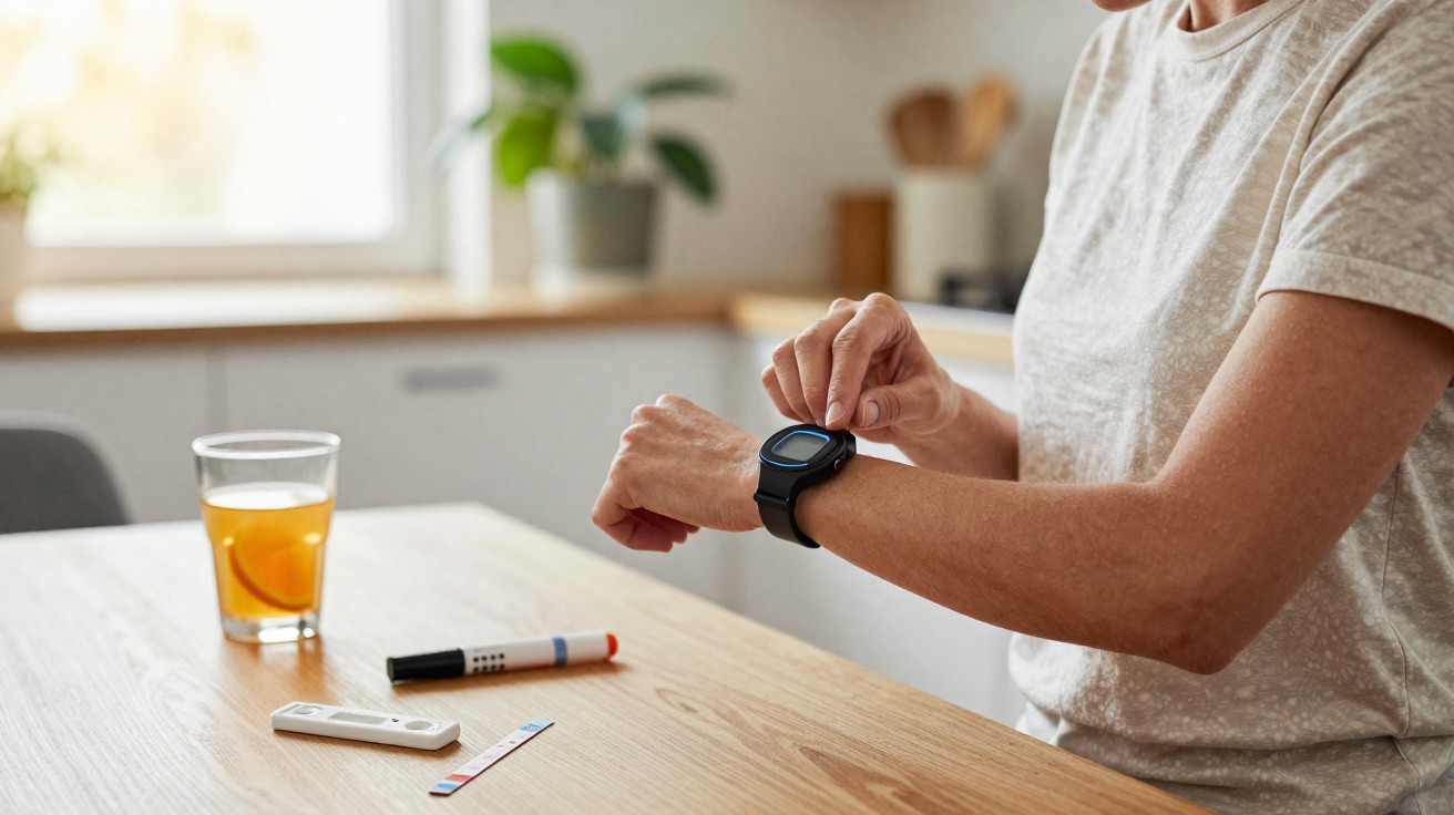
A familiar scene plays out in millions of homes: the alarm sounds, someone takes a deep breath, readies the lancing device and still feels that slight fear of pain.
The routine of managing diabetes remains demanding, even with modern sensors attached to the arm. Researchers at MIT now say they have taken a practical step towards changing this situation with a technology that reads glucose without piercing the skin, using only beams of light.
A small action with a huge burden
Anyone living with diabetes knows that management involves more than medication. It begins at the fingertip, several times a day, every day.
Every finger prick causes physical discomfort, but it also carries an emotional burden. After years of doing it, many people begin avoiding tests, cutting corners or missing scheduled checks. Guilt and fear of complications can follow, along with, in many cases, a quiet withdrawal from their own care.
Even subcutaneous sensors, widely seen as an advance, have barriers of their own. They must be inserted with a specialised needle, may irritate the skin, require regular replacement and are too expensive for some people. In many countries, consistent access is not available to everyone.
“The biggest challenge today is not simply measuring glucose accurately, but making it so simple and painless that patients do not hesitate before looking after themselves.”
When monitoring breaks down, glucose levels can fluctuate without control. Persistent tiredness, gradual loss of vision, and kidney and heart problems are only some of the potential consequences. That is why laboratories around the world are pursuing a reliable needle-free solution.
Light instead of a needle: what MIT is testing
Researchers at MIT in the United States have chosen a less obvious route: using light to ‘see’ glucose beneath the skin, without cutting, piercing or placing anything inside the body.
The technology relies on Raman spectroscopy. Put simply, it examines the way light interacts with molecules in the body. When a beam reaches the skin, some of that light is scattered in a distinctive manner, creating a kind of ‘fingerprint’ for the substances present.
For glucose, the device directs near-infrared light at the forearm. The detected signal comes mainly from interstitial fluid, the fluid located between cells just below the skin’s surface. This fluid closely follows changes in blood glucose.
From a printer-sized machine to a tabletop device
The idea is not new. Since 2010, the MIT team has shown that Raman spectroscopy could detect glucose without invasive contact. The issue was scale: the earliest systems took up as much room as a large printer and contained numerous complex optical components.
In recent years, the researchers have managed to reduce the system to something roughly the size of a shoebox, without sacrificing as much accuracy. Their approach was to select just three highly specific spectral bands from thousands of possible light ranges, each linked to glucose behaviour.
“By concentrating on three very clearly defined light windows, MIT reduced cost, space and processing time while keeping the glucose signal at the centre of the analysis.”
Each reading takes around 30 seconds. In initial tests involving one healthy volunteer, the findings were compared with two systems already established on the market: the Freestyle Libre 3 and Dexcom G7, both of which use sensors beneath the skin. The optical technology achieved similar levels of accuracy, which is unusual among entirely non-invasive methods.
How the new BRS prototype works
In the latest publication, the device was given a name: BRS, the English abbreviation for “band-restricted Raman spectroscopy”. It was built into a housing measuring approximately 31 × 27 × 21 cm.
In this version, the device focuses on three specific wavelengths:
- one central band, aligned with the strongest glucose signal;
- two side bands, which serve as internal references to correct for noise and skin variation.
During testing, an 830 nm beam, within the near-infrared range, was directed at one participant’s forearm every five minutes for four consecutive hours. At the same time, measurements were taken using a conventional blood glucose meter and subcutaneous sensors.
The data were processed with a quadratic calibration algorithm, designed to connect the captured light pattern with the actual glucose value. The mean relative error was around 12%, a level regarded as acceptable for clinical applications, particularly at an experimental stage.
From a shoebox to the patient’s wrist
The next step announced by the researchers is more ambitious still: shrinking the system to the size of a wristwatch or a small wristband. This would be the stage at which the device moves beyond the laboratory and closer to an everyday product.
Tests have already begun with people with pre-diabetes and with different skin tones, an essential consideration. Skin can significantly affect its interaction with light, and the aim is to ensure consistent performance across varied user profiles.
“If miniaturisation preserves accuracy, checking glucose could become as simple as looking at the time on a watch.”
What this could change for people with diabetes
A dependable optical sensor could reshape a patient’s relationship with treatment. With no pain or blood involved, testing is likely to happen more often. More data increases the opportunity for fine adjustments to diet, insulin and physical activity.
In a practical setting, a wristband-shaped device could send alerts when glucose began rising or falling too quickly, even before symptoms of hypoglycaemia or hyperglycaemia appeared. This could reduce hospital admissions and severe episodes while providing a greater feeling of security.
For doctors and healthcare teams, a monitor of this kind would create a detailed record of fluctuations through the day and night. That would make it easier to alter treatment plans, identify the most critical times and make personalised adjustments.
Limitations, risks and realistic expectations
Needle-free technologies often generate considerable excitement, but history includes many promising projects that were eventually shelved. Insufficient accuracy, high costs and difficulties with large-scale production are recurring obstacles.
For the MIT device, several questions remain unresolved:
- how the device will cope with sweat, body hair, tattoos or creams on the skin;
- the effect of changes in ambient temperature on readings;
- the durability of optical components during everyday use;
- the final cost to users or healthcare systems.
There is also a risk of false reassurance: if the sensor consistently produces readings that are slightly removed from reality, decisions about insulin doses could be compromised. Before reaching the market, the technology must undergo studies involving hundreds or thousands of people in different countries and settings.
Terms worth a quick explanation
| Term | What it means |
|---|---|
| Interstitial fluid | Fluid that fills the spaces between cells and contains glucose at levels close to those in the blood. |
| Raman spectroscopy | A technique that analyses how light is scattered by molecules to identify substances in a material. |
| Calibration algorithm | A set of calculations that converts the captured optical signal into numerical glucose values. |
| Continuous glucose monitor | A sensor that measures glucose levels at regular intervals, providing a near-real-time graph. |
Possible scenarios over the next few years
If MIT’s technology progresses as planned, different combinations of diabetes care are conceivable. A patient could wear an optical wristband for continuous monitoring and turn to a conventional blood glucose meter only to confirm values in particular situations.
Health insurers and public health systems may come to regard this type of device as a strategic investment: fewer serious complications mean fewer expensive hospital admissions in the future. For people with pre-diabetes, a painless sensor could act as a motivational tool, helping them follow the direct effect that changes in diet and exercise have on glucose.
At the same time, success with this method could create scope for applying the same principle to other markers in the body. Cholesterol, hormones and inflammation markers: anything that interacts clearly with light becomes a research target. Gradually, the human body could become more ‘readable’ without cuts or needles, simply through discreet sensors used throughout everyday life.






Comments
No comments yet. Be the first to comment!
Leave a Comment